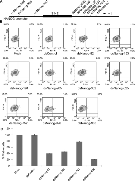
Figure 1. Identification of NANOG saRNAs using a GFP reporter system.
(A) Schematic representation of the NANOG promoter. Indicated are the names of each saRNA and its target site relative to the TSS (+1). A SINE (short interspersed element) repetitive element is also indicated in the NANOG promoter. (B) NCCIT cells were infected with lentiviral particles containing GFP under the control of the human NANOG promoter. At 3 days after transduction GFP-positive cells (NCCIT NANOG–GFP) were collected by cell sorting and subcultured for saRNA transfection. NCCIT NANOG–GFP cells were transfected with 50 nM concentrations of the indicated saRNAs for three days and analysed by FACS to assess GFP intensity in each population. Mock samples were transfected in the absence of saRNA. Representative data of n=2. (C) NCCIT cells were transfected with the indicated dsRNA at 100 nM. MTT assay was conducted 96 hours following transfection. Results are plotted as the percentage of viable cells relative to mock transfections (means±S.D. for two independent experiments).

