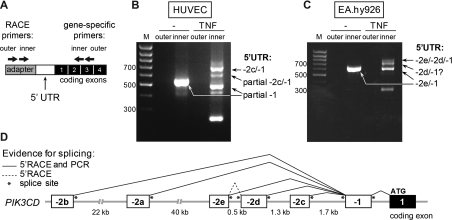
Figure 2. Novel TNFα-inducible PIK3CD TSSs uncovered by 5′RACE in human ECs.
(A) Schematic representation of the 5′RACE strategy used to analyse the PIK3CD 5′-UTR in human ECs. The 5′RACE adapter is shown in grey, the 5′-UTR of the human PIK3CD mRNA transcript is shown in white and the protein-coding exons are shown in black. The approximate binding sites of the outer and inner primers used in the nested PCR are depicted with arrows. (B and C) 5′RACE was performed in unstimulated and TNFα-stimulated (10 ng/ml for 18 h) HUVECs (B) and EA.hy926 cells (C). Nested PCR was performed on 5′RACE cDNA using the outer and inner primer pairs shown in (A) and products of both reactions were analysed on a 1% agarose gel. Arrows indicate the cloned and sequenced RACE products. In unstimulated HUVECs, the 5′-UTR of p110δ transcripts contains a partial exon −1 spliced on to exon 1 without a −2 exon and in TNFα-stimulated HUVECs, exon −2c spliced on to the full-length exon−1. In both unstimulated and TNFα-stimulated EA.hy926 cells, the 5′-UTR of p110δ transcripts is formed of exon −2e spliced on to full-length exon −1 or exon −2d spliced between exons −2e and −1. No 5′RACE products with exon−2d as their first 5′ exon were found although a band with an estimated molecular mass corresponding to such a 5′RACE product is visible on the agarose gel in TNFα-stimulated EA.hy926 cells. The additional visible bands are degradation products containing partial sequences of exons −2c or −1. (D) Schematic representation of human PIK3CD 5′ untranslated exons and their splicing pattern as identified by 5′RACE or by PCR amplification. M, molecular mass marker.

