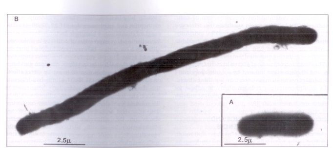
Figure 1.
Electron micrograph showing A an Escherichia coli growing in Mueller-Hinton broth and B a filamentous E coli as a result of exposure to ampicillin at one-half minimal inhibitory concentration for 2 h. Cells were stained with 2.5 mmol phosphotungstic acid adjusted to pH 7.0 with sodium hydroxide, and viewed on a Phillips model 201 electron microscope (magnification ×9000)

