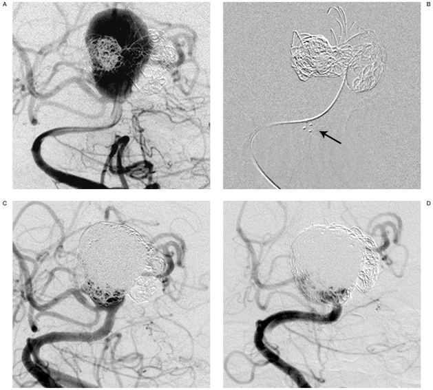
Figure 1.
A) Anteroposterior view of the right vertebral artery injection demonstrates a recurrent basilar bifurcation aneurysm incorporating both posterior cerebral arteries and the right superior cerebellar artery. B) Anteroposterior angiographic view shows the aneurysm with platinum Neuroform stent marks in the basilar trunk (arrow). Distal marks in the fundus of the aneurysm are obliterated by the coil mass. C) Anteroposterior angiogram shows near-complete coiling of the aneurysm. D) Six-month follow-up anteroposterior angiography shows persistent occlusion of the aneurysm.

