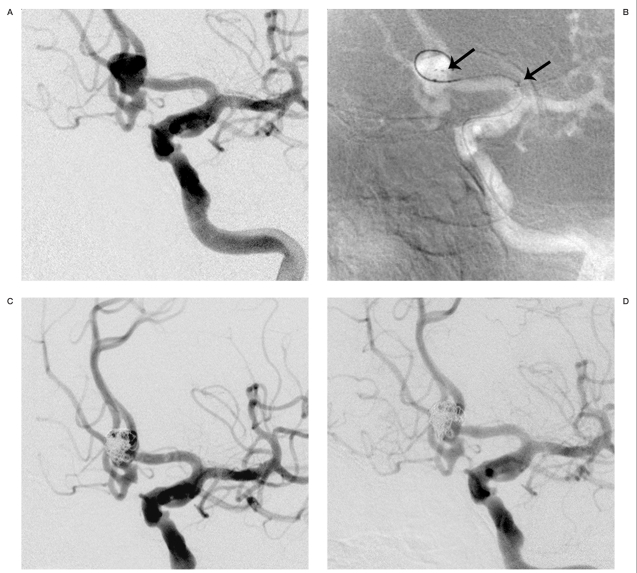
Figure 2.
A) Anteroposterior view of the left internal carotid artery injection demonstrates a 7-mm wide-necked anterior communicating artery aneurysm incorporating A2 segments of the bilateral anterior cerebral arteries. B) Anteroposterior angiographic view shows the aneurysm with platinum Neuroform stent marks in the fundus and in the A1 segment of the left anterior cerebral artery (arrows). C) Anteroposterior angiogram shows the completely coiled aneurysm. D) Six-month follow-up anteroposterior angiography shows persistent occlusion of the aneurysm.

