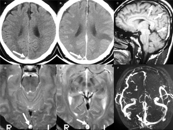
Figure 1.
The intravascular thrombus (arrow) within the superior sagittal sinus is elucidated as hyperdensity on non-contrast CT (A), the triangular defect (empty delta sign) on contrast enhancing CT (B), heterogeneous hyperintensity on sagittal T1WI (C) and axial T2WI(D,E) MR images. The 2D TOF MRV shows absence of right transverse sinus with incomplete filling of right sinus confluence (F).

