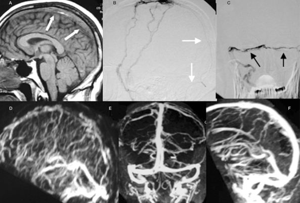
Figure 4.
The T1 weighted MR image (A) shows diffuse intraluminal thrombus within the superior sagittal sinus (arrow). During retrograde transvenous thrombolytic treatment, the micro-catheter (arrow) is demonstrated on lateral view (B) with the presence of filling defects caused by the thrombi (black arrow) within both transverse sinuses (C). The patient received subsequent heparinization for 4 days and was improved clinically. The follow-up PA (E) and lateral (F) view of the CEMRV at two weeks shows a more patent venous sinus when compared to the lateral view of MR venography done the next day of treatment (D).

