Figure 3.
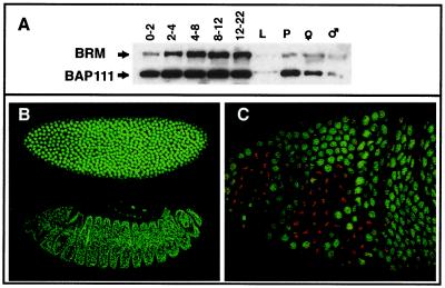
Developmental expression of the BAP111 protein. (A) Ten micrograms of protein extracted from staged embryos (0–2, 2–4, 4–8, 8–12, and 12–22 h), larvae (L), pupae (P), and adult females (♀) or males (♂) was analyzed by Western blotting with anti-BRM and anti-BAP111 antibodies. Blots are overexposed to reveal the signal in larvae. (B) Affinity-purified anti-BAP111 antibodies detect ubiquitous nuclear staining of BAP111 in syncytial blastoderm embryos (Upper) and germ band retracted embryos (Lower). (C) BAP111 is not associated with mitotic chromosomes. Higher magnification (630×) view of a cephalic-furrow stage embryo stained with BAP111 antibodies (green) and propidium iodide to visualize DNA (red). BAP111 is nuclear except in domains of mitotic activity where BAP111 becomes dispersed throughout the cell.

