FIG. 3.
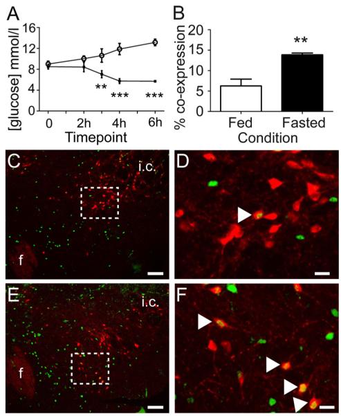
Acute food deprivation decreases blood glucose and increases FOS-IR in LHA NPY neurons. A, Mice that underwent a 6-h fast from 1600 to 2200 h demonstrated significantly lower blood glucose compared with ad libitum-fed controls (open circles, ad libitum fed; black squares, fasted). B, A separate group of mice that underwent a 6-h fast from 1600 to 2200 h demonstrated significantly more FOS-IR within GFP-IR neurons of the LHA compared with ad libitum-fed control animals. Representative images (C–F) showing FOS-IR (green) and GFP-IR (red) within the LHA of ad libitum-fed (C, low magnification; D, high magnification) or 6-h fasted (E, low magnification; F, high magnification) mice. Boxed areas indicate the regions shown at high magnification. White arrowheads in D and F indicate double-labeled neurons. F, Fornix; i.c., internal capsule. Scale bars (C and E), 100 μm; (D and F), 20 μm. **, P ≤ 0.01; ***, P ≤ 0.001.

