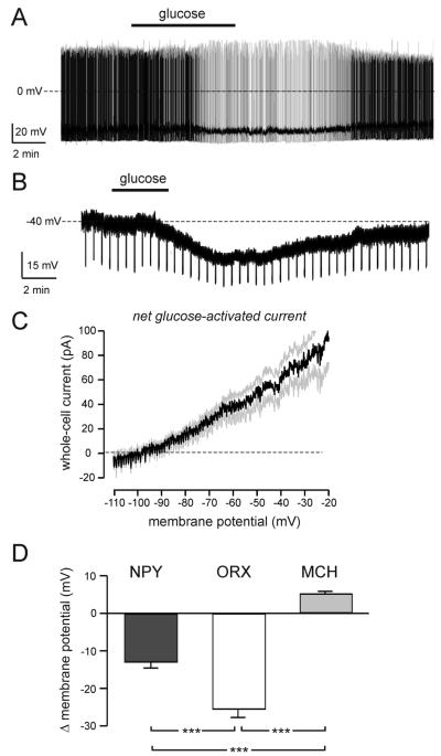
FIG. 5.
Glucose sensing by LHA NPY neurons in situ. A, Typical effect of an increase in extracellular glucose (switch from 1 to 5 mM glucose marked with black bar) in a spontaneously firing LHA NPY-GFP cell. Similar responses were obtained in seven cells. B, Effect of glucose (same concentration switch as in A) on the membrane potential of an LHA NPY-GFP cell in the presence of 1 μM tetrodotoxin. Downward deflections are responses to fixed-amplitude, 1-sec-long injections of hyperpolarizing current (note that elevated glucose decreases their amplitude, implying an increase in membrane conductance). Similar responses were obtained in six cells. C, The current-voltage relationship of the net glucose-activated current. Black line represents mean, gray lines represent SEM (means ± SEM of six cells). D, Effect of increased extracellular glucose (1 to 5 mM switch) on the membrane potential of LHA NPY (n = 6), MCH (n = 14), and ORX (n = 14) cells, recorded in the presence of 1 μM tetrodotoxin. ***, P ≤ 0.001.

