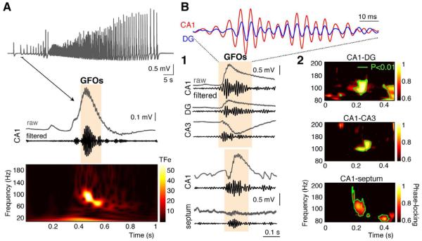
Figure 1. GFOs at seizure onset in the immature SHF.
(A) ILE recorded extracellularly in the CA1 area of a P7 SHF expresses GFOs at its onset as evidenced by 60-120 Hz band-pass filtering and by the time-frequency (TF) representation (TFe: TF energy, color coded in standard deviations above the mean power of an event-free baseline).
(B1) GFOs occurs simultaneously in the dentate gyrus (DG) and in the CA3 and CA1 regions, as well as between the septum and the CA1 region.
(B2) TF analysis of phase-locking between the different regions of the SHF reveals a significant enhancement of phase-synchrony around 100Hz (green contour line: p<0.01).

