Fig. 6.
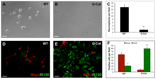
β-catenin-expressing stem cells exhibit aberrant self-renewal and differentiation in vitro. (A,B) Prom1+ Lin– cells were sorted from P0 WT (A) or G-Cat (B) mouse cerebellum and cultured at clonal density in serum-free medium containing 25 ng/ml EGF and bFGF for 7 days before being photographed under bright-field. Passageable neurospheres were consistently obtained from WT but not from mutant cerebellum. (C) Average neurosphere numbers (±s.e.m.) per field from eight fields in P0 WT or G-Cat cerebellum. (D,E) Prom1+ Lin– cells were sorted from P0 WT (D) or G-Cat (E) cerebellum and cultured for 3 days in medium containing 1% FBS. Cells were stained with antibodies specific for the neuronal marker Map2 (red) and the glial cell marker S100β (green). (F) Quantitation (mean ± s.e.m.) of Map2/S100β staining from five fields. *, P<0.01; **, P<0.001. Whereas WT stem cells differentiated predominantly into neurons, G-Cat showed a marked skewing toward glial lineages. Scale bars: 25 μm in A,B; 50 μm in D,E.

