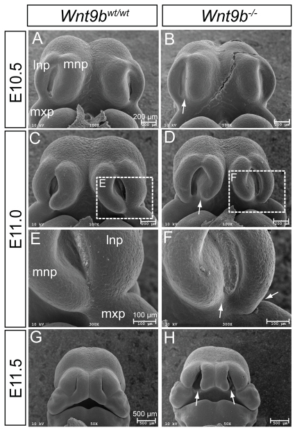
Fig. 2.
Scanning electron microscopy analysis of facial development. (A-H) Front facial views of wild-type (A,C,E,G) and Wnt9b–/– (B,D,F,H) mouse embryos from E10.5 to E11.5. The boxed regions in C and D are magnified in E and F, respectively. Reduced invagination between the MNP and LNP was clear in Wnt9b–/– embryos at E10.5 (arrow in B). At E11.0, hypoplasia of the MNP and LNP was obvious, and the MNP, LNP and MxP failed to complete fusion (arrows in D,F). At E11.5, bilateral lip clefts (arrows in H) were apparent in Wnt9b–/– embryos. lnp, lateral nasal process; mnp, medial nasal process; mxp, maxillary process.

