Fig. 3.
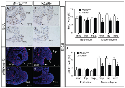
Cell proliferation in the lip fusion zone. (A-D) Immunostaining for incorporated BrdU in the facial processes at E10.5. Note that the epithelial layer in the fusion area lacks BrdU-positive cells (arrows). (E-H) Mitotic cells detected by phospho-histone H3 in the facial processes at E10.5. The epithelial layer in the fusion area lacks phospho-histone H3-positive cells (arrows). (I,J) For calculating the proliferation rate, the numbers of BrdU-positive or phospho-histone H3-positive cells were counted in each of two sections of three embryos. Mean±s.e.m. *P<0.05, Student’s t-test. lnp, lateral nasal process; mnp, medial nasal process; mxp, maxillary process.

