
Figure 3.
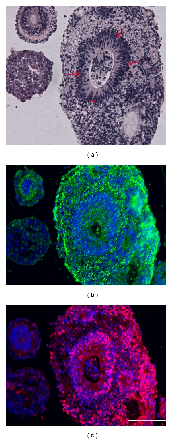
Immunohistochemical analysis of embryoid body sections confirming neuroepithelial tissue in adjacent sections, hematoxylin, and eosin-stained neural rosettes in hiPSCs-derived EBs (a), antinestin immunofluorescent staining (b), green-nestin, blue-nuclei stain, and anti-Sox2 immunofluorescent staining (c), red-Sox2, blue-nuclei stain. Magnification is 400x total (10x ocular, 40x objective). Each scale bar represents 50 μm in length.