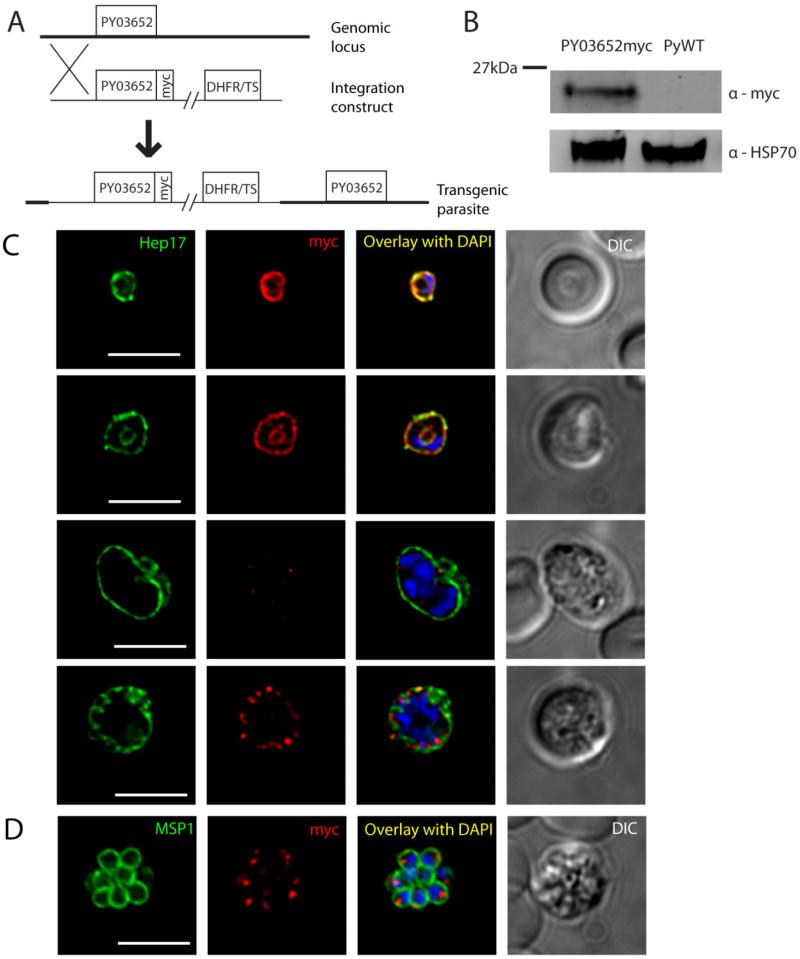
Figure 3.
Protein expression and localization of PY03652myc in blood stages. (A) Schematic representation of the strategy used to insert a construct encoding a c-terminal fusion of the 4x myc epitope to PY03652 upstream of the endogenous PY03652 ORF. (B) Western blot of lysates from mouse blood infected with asynchronous PY03652myc parasites or WT control. (C) IFA images of PY03652myc blood stages labeled with anti-myc or anti-Hep17 antibodies, and DAPI (to label DNA). A differential interference contrast (DIC) image of the liver section is shown to the right. Overlap of myc signal with Hep17 indicates localization of PY03652myc to the PVM in ring and trophozoite stages. Myc staining was weak or absent in early schizonts, but appears again in late schizogony, when it localizes to vesicular structures in the parasite periphery, not in the PVM. Scale bars = 5 micrometers. (D) IFA of blood stage PY03652myc schizont, labeled with anti-myc or anti-MSP1 antibodies, and DAPI. The myc signal appears in small foci within the daughter merozoites, likely within secretory organelles. Scale bar = 5 micrometers.

