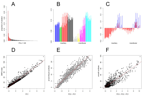
Figure 1.
Principal component analysis. (A) proportion of data variance explained by PCs 1-10 (red) and successive PCs (black). (B) Loadings for PC1 ordered by tooth type, from left to right: maxillary incisors (blue), canines (green), premolars (red), molars (black), mandibular incisors (gray), canines (yellow), premolars (magenta), molars (cyan). For each tooth, contributions of surfaces are listed in the following order: buccal, distal, lingual, mesial, and occlusal, if applicable. Teeth ordered from left to right: maxillary 8, 9, 7, 10, 6, 11, 5, 12, 4, 13, 3, 14, 2, 15; mandibular 24, 25, 23, 26, 22, 27, 21, 28, 20, 29, 19, 30, 18, 31. (C) Loadings for PC2, in the same order; smooth surfaces shaded red, pit and fissure surfaces shaded blue. Scatter plots of (D) PC1 vs. DMFS index, (E) PC2 vs. pit and fissure caries, (F) PC3 vs. smooth surface caries.

