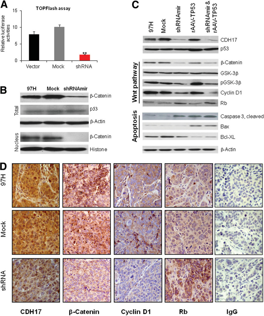
Fig. 6.
Suppression of the β-catenin/Wnt pathway in MHCC97H cells in vitro and in vivo by knockdown of CDH17. (A) A TOPFlash reporter luciferase assay was performed using MHCC97H cells transfected with (shRNA) or without (Vector and Mock) CDH17 shRNA. Luciferase intensities for each experimental group were quantified using a luminometer. The relative luciferase activities were calculated when the luciferase intensities of the pRL-TK-Luc vector were taken into account. **P < 0.0001. (B) Western blots of β-catenin and p53 in MHCC97H cells infected with (shRNAmir) or without (97H and Mock) CDH17 shRNAmir. (C) For the in vivo study, subcutaneous tumors were induced in nude mice and CDH17- and p53-associated gene therapies were applied to the tumors directly. The effects on the expression of a panel of proteins associated with the Wnt pathway and apoptosis were revealed by way of western blotting for each treatment group. (D) Tumors were subsequently dissected, then stained immuno-histochemically using antibodies specific for CDH17, β-catenin, cyclin D1, and Rb proteins. Immunoglobulin G was used to substitute the antibody to perform the immunohistochemistry to reveal the authenticity of the staining. (Original magnification ×400.)

