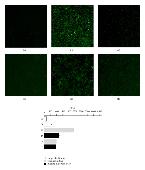
Figure 4.

The cell imaging and fluorescence intensity reflecting the interactions between soluble DC-SIGN receptor with the LeX and LeY determinants exposed on the surface THP-1 leukemia cells, in the immunofluorescence assay. Control THP-1 cells, nondifferentiated (a) or differentiated (b) with PMA, GM-CSF and IL-4 stained with FITC-conjugated sheep antibodies against mouse Igs (secondary Ab). Differentiated THP-1 cells treated with anti-LeX mAb and FITC-conjugated secondary Ab (c) or firstly treated with soluble DC-SIGN followed by treatment as above (d). Differentiated THP-1 cells treated with anti-LeY mAb and FITC-conjugated secondary Ab (e) or firstly treated with soluble DC-SIGN followed by the treatment as above (f).
