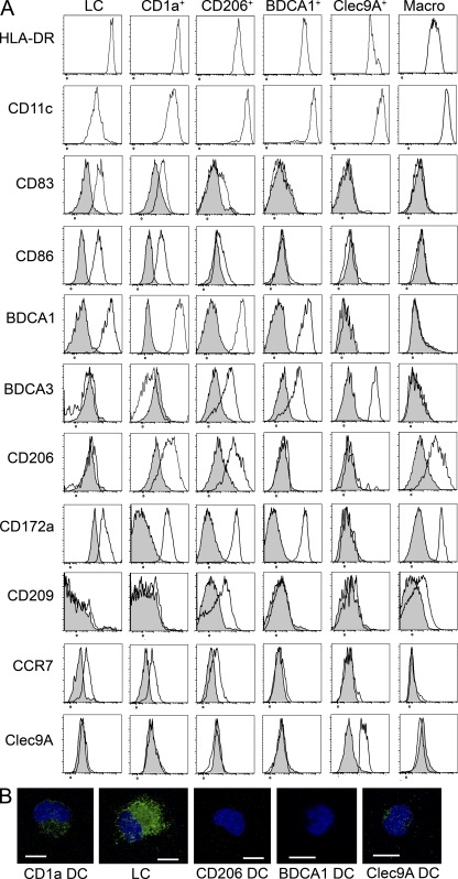
Figure 3.
Phenotype of LN DC subsets. (A) Axillary LN cells were enriched for DCs and stained for HLA-DR, CD11c, CD1a, CD14, EpCAM, BDCA1, Clec9A, CD206, CD83, CD86, BDCA3, CD172a, CD209, CCR7, or control isotype. Representative results of 4–12 independent experiments are shown. (B) Purified DC populations were coated on microscopy slides, fixed, permeabilized, and stained for langerin (green). The nucleus is stained with DAPI (blue). Representative images are shown of three to four independent experiments. Bars, 10 µm.

