Figure 6.
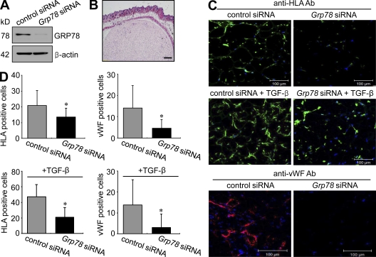
Inhibition of RA synoviocyte proliferation and angiogenesis by Grp78 siRNA in immunodeficient mice. Matrigel containing RA FLSs was subcutaneously injected into the back skin of immunodeficient mice. (A) 7 d after transfecting with the Grp78 siRNA, GRP78 expression levels in RA FLSs were determined by Western blot analysis. (B) Hematoxylin and eosin staining of Matrigels containing RA FLSs from immunodeficient mice. (C) Effect of Grp78 siRNA on RA FLS proliferation and HUVEC infiltration. RA FLSs were implanted in Matrigels for 7 d in the absence or presence of 50 ng/gel TGF-β. RA FLSs in Matrigel were identified by immunofluorescence labeling for HLA class I antigen. Infiltrating mouse endothelial cells were stained using mouse anti-vWF antibody. The cells positive for HLA class I are shown in green in the top and the middle panels. Representative photographs of endothelial cells are shown in red in the bottom panel. Bars: (B) 300 µm; (C) 100 µm. (D) Numbers of RA FLSs and endothelial cells in Matrigels treated with vehicle or TGF-β. Cells were manually counted under a magnification of ×200. Values are the mean ± SD of eight mice per group. *, P < 0.05 versus control siRNA–transfected cells.

