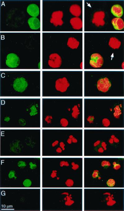
Figure 6.
The nuclear lamina in K562 cells is disrupted after PFP-loading Gzms. K562 cells, mock-treated (A), or treated with PFP alone (B) or with PFP and GzmA (C–E) or GzmB (F and G) for 1 h, were stained with FITC-anti-laminB mAb 101-B7 (Left) or propidium iodide (Center); overlay (Right). Arrows in A and B indicate dissolution of the lamina and dispersion of laminB to the cytoplasm in mitotic cells. (C–E) Progressive changes in laminB degradation after GzmA treatment. Before chromatin changes are evident (C), lamin staining becomes punctate. As chromatin condensation and nuclear fragmentation progress, the nuclear lamina dissolves and lamin staining diminishes and disperses to the cytoplasm. K562 cells loaded with GzmB exhibit less pronounced changes in laminB staining. These changes persist even with CI (G).

