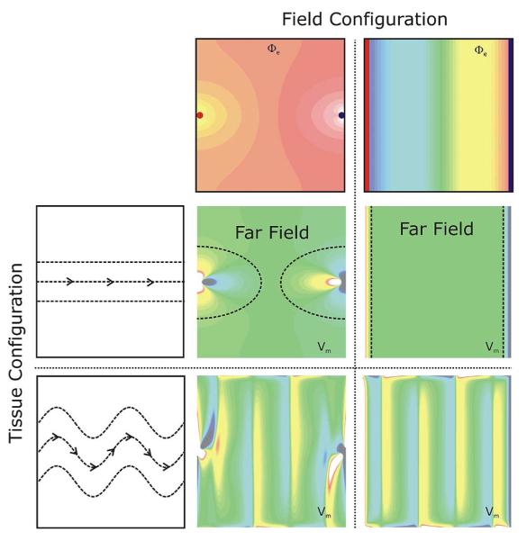
Fig. 5.
An important insight learned from the bidomain model is that the etiology of VEPs is determined by both field configuration as well as tissue structure. Shown are shock-induced polarization patterns as a function of field and tissue configuration. Top panels: Extracellular potentials Φe, as induced by two point (left) and line electrodes (right). Red (blue) indicates cathodal (anodal) stimulus. Left panels: Two tissue structure configurations are shown, a homogeneous configuration with straight fibers (top), and a configuration where fiber orientation varies as a function of space (bottom). Fiber orientation is indicated by the dashed lines. Central panels: Shock-induced polarization patterns for all possible combinations between field and tissue configuration. In the case of plate electrodes with a homogeneous tissue structure only surface polarizations close to the electrode locations are observed; the bulk of the tissue remains essentially unaffected.

