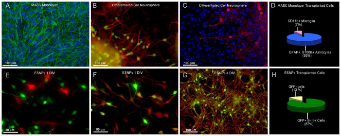
Figure 1. In vitro characterization of both donor populations shows contrasting cell types.
A) Cerebellum-derived MASCs derived from p0–p8 postnatal mouse cerebellum and cultured as GFAP+ (green) astrocyte monolayers were used for in vivo transplantation. (blue=DAPI) B) When cultured as neurospheres, MASCs are capable of differentiating into neurons expressing neuronal markers including β-III tubulin (red) and NeuN (green). C) MASC-derived neurospheres are also found to contain CNPase (red)-expressing oligodendrocytes. (blue=DAPI) D) Quantitative analysis shows that MASC monolayers used for transplantation contain mostly astrocytes (93% as shown in blue) and a few microglia (7% as shown in pink). τEGFP-ESNP cells, on the other hand, express mostly a neuronal fate in culture. E) Following one day in culture, GFP+ ESNPs (green) are not immunopositive for GFAP (red), however, F) shows that they do co-localize with the neuronal marker β-III tubulin (red). G) ESNPs used for in vivo transplantation are mostly immunolabeled for both GFP (green) and β-III tubulin (red) after four days in culture. H) Quantitative analysis shows that ESNPs contain mostly GFP+ immature neurons (87% as shown in green). (DIV=days in vitro)

