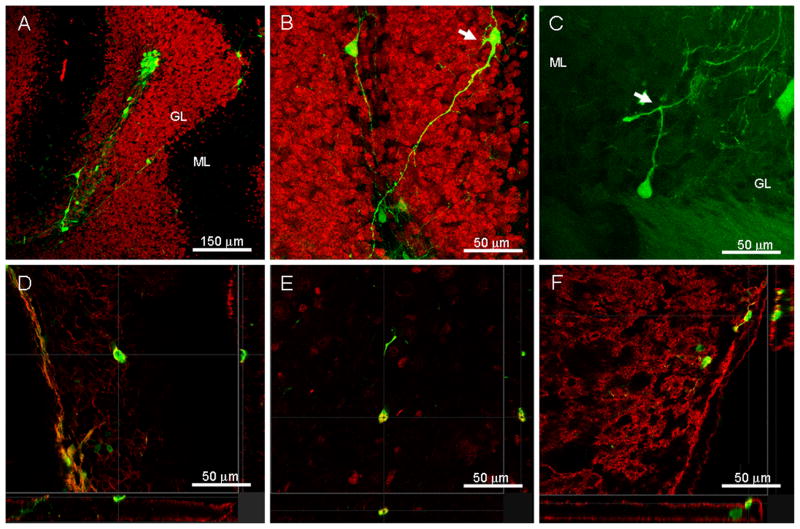
Figure 3. Grafted ESNPs have the ability to survive, migrate, and differentiate into mature cellular phenotypes following transplantation into the weaver mouse cerebellum.
A) Confocal microscopy shows that grafted GFP+ ESNPs (green) can re-aggregate into cell clusters, but they retain the ability to migrate away from the injection site. B) Higher magnification image shows that some of these cells display mature neuronal phenotypes (arrow) with ramified processes and thin, varicose axons. C) An example of a small number of the donor-derived cells (green) found to possess axon-like processes that bifurcate into T shapes (arrow), resembling parallel fibers of the granule neurons. D) A small population of the donor cells also express the pan neuronal marker β-III tubulin (red), the mature neuronal marker NeuN (red) in E), and in F) the neurotransmitter glutamate (red) which is expressed exclusively by granule cells within the cerebellum. (GL=granule layer, ML=molecular layer)

