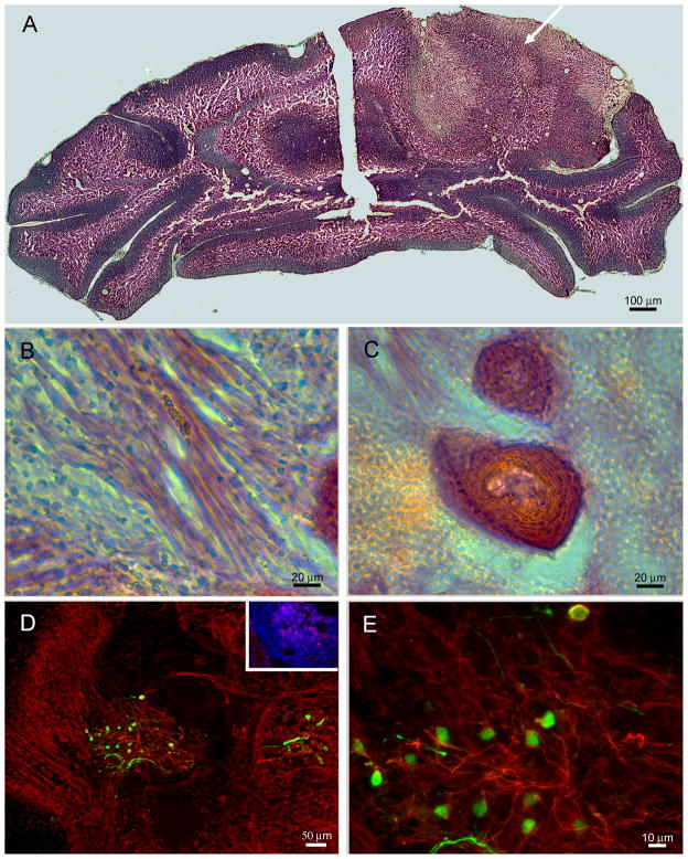
Figure 4. Grafted ESNPs are capable of transforming host tissue and give rise to tumor-like spheres.
A) Montage of images from H& E staining shows the transplanted hemisphere being disrupted by a protruding cell mass (arrow) within the parenchyma but not on the control contralateral side. B) Non-neural lineages such as skeletal muscle fibers or in C) hair follicles can be seen with the teratoma-like neoplasia within the cerebellum based on morphological analysis from H&E staining of the tissue. D) GFP+ ESNPs (green) can be seen within the transformed tissue, and they are also immunopositive for SSEA-1 (red, inset), and β-III tubulin (red) in D and E. Presence of GFP+, β-III tubulin+ cells, same as the grafted population, within the transformed tissue suggests the ESNPs are the most probable cells of origin for the tumor-like neoplasias.

