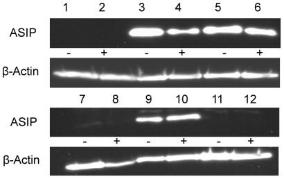
Figure 5. ASIP protein expression in different tissues of bulls with normal expression (−) or over-expression of ASIP mRNA (+).
Chemiluminescence detection of ASIP and β-actin by Western blotting of 40 µg protein of the respective tissues. Lanes 1 and 2: M. longissimus, 3 and 4: subcutaneous fat, 5 and 6: intermuscular fat, 7 and 8: heart, 9 and 10: liver, 11 and 12: lung.

