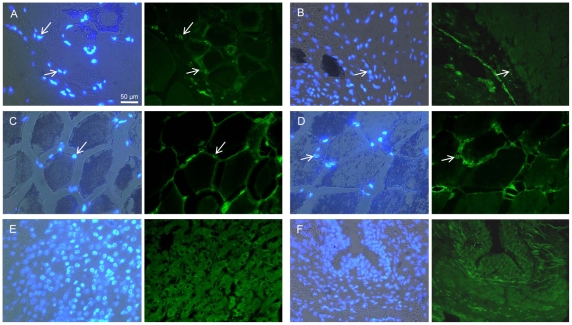
Figure 6. Cellular localization of ASIP protein in different bovine tissues.
The left panels show the overlay of Hoechst 33258 nuclear stain with the bright-field image. In the right panels, ASIP protein is labeled with Alexa 488 (green). White arrows indicate distinct signals located at nuclei. (A) M. longissimus with included intramuscular fat. (B) cardiac muscle, (C) subcutaneous fat, (D) intermuscular fat, (E) liver, (F) lung.

