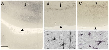
Figure 5. Histological staining for iron with Perl's DAB stain in the motor cortex.
The panels show comparable planes of section through the precentral gyrus of brains from the second ALS patient (A), and from the Alzheimer (B) and Parkinson (C) patients. The arrowheads indicate the pial surface. The arrows indicate the gray-white junction. (A) Low power magnification showing iron accumulation in the middle and deeper layers of cortical gray matter, and at the gray-white junction in a 51-year-old ALS patient. At higher power (D, E) the staining is present in cells with irregular processes suggestive of microglia in the ALS motor cortex. The lower portion of panel A shows the post-central gyrus with relatively little iron staining. The iron staining was more diffuse within the motor cortex of Alzheimer (B) and Parkinson (C) patients and was also present in the subcortical white matter. Scale bars, 1 mm (A–C), 10 µm (D, E).

