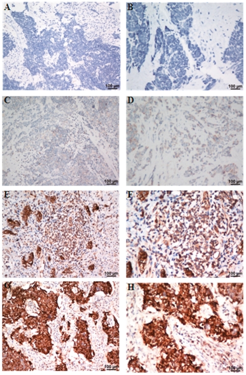
Figure 2. 2. Immunohistochemical analyses of chromogranin A (CgA) staining in Small cell carcinoma of the cervix tissues.
As can be seen, CgA shows no positive staining (−)(case-431901; A: x200; B: x400), weakly positive staining (+) (case-385737; C: x200; D: x400), moderate staining (++) (case-449570; E: x200; F: x400), strong staining (+++) (case-437412; G: x200; H: x400) in Small cell carcinoma of the cervix tissues.

