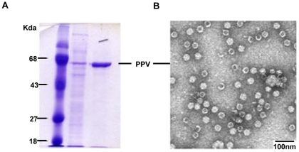
Figure 1. Characterization of PPV-PYCS VLPs.
Conditions for generation of the PPV vector expressing the specific CD8+ T cell epitope of P. yoelii CS, growth in baculovirus-infected insect cells and purification of the PPV-VLPs is described under Materials and Methods. (A). Proteins were resolved by 9% SDS-PAGE and visualized after coomassie-blue staining. Molecular masses of standard proteins (lane 1) are indicated at the left. Lane 2, shows partial purification and lane 3, purified protein with the size corresponding to the PPV VP2. (B). Electron microscope image of negatively stained PPV-PYCS VLPs.

