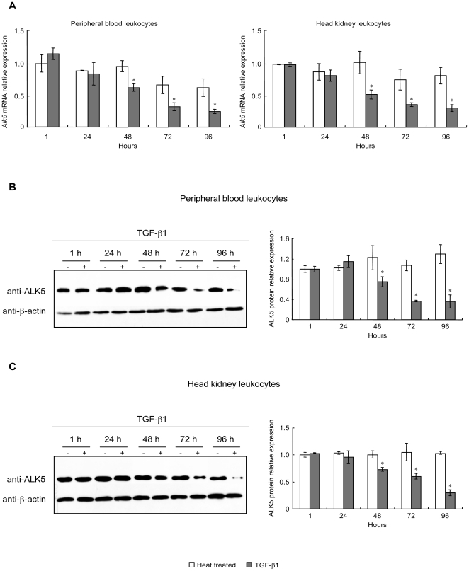
Figure 4. Effects of TGF-β1 on ALK5 mRNA and protein levels in PBL and HKL.
After treatment with native or heat treated rgcTGF-β1 (100 ng/ml) for 1–96 h, ALK5 mRNA (A) and protein (B–C) levels in PBL or HKL were analyzed by qPCR and WB, respectively. In parallel experiments, leukocytes were exposed to the decreasing dilution of gcTGF-β1 mAb (1∶30000-1∶300) for 72 h under static incubation. Subsequently, qPCR and WB were performed for detection of ALK5 mRNA expression (D) and protein levels (E) in PBL (left panels) or HKL (right panels). Relative mRNA expression levels of Alk5 were analyzed using β-actin as an internal reference and expressed as the fold changes of the heat treated group at 1 h. Data presented (mean±SEM, N = 4) are representative results from three individual experiments. In WB, the representative results were showed here and β-actin levels were used as an internal control and isotype control was mouse IgG (30 µg/ml). Meanwhile, the densitometric analysis of ALK5 protein levels was performed (mean±SEM, N = 4) and the relative protein levels were expressed as the fold changes of the heat treated group at 1 h. The asterisk (*) denotes a significant difference at P<0.05.

