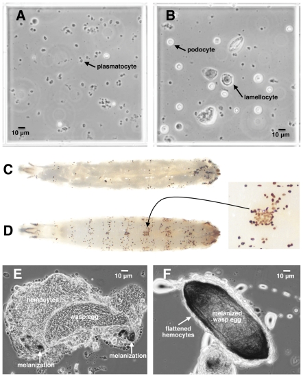
Figure 3. D. suzukii hemocytes and encapsulation of wasp eggs.
(A) A 0.25×0.25×0.1 mm hemocytometer field from normal D. suzukii larvae showing abundant plasmatocytes; (B) hemocytometer field from D. suzukii larvae 12 hours after infection by wasp strain LbG486 showing increased podocyte and lamellocyte numbers; (C) control D. melanogaster larva with melanized crystal cells; (D) control D. suzukii larva with melanized crystal cells, showing color variation in inset; (E) initiation of encapsulation of LbG486 egg by D. suzukii showing loose hemocyte aggregation and melanization at anterior and posterior tips of egg; (F) LbG486 egg melanotically encapsulated by D. suzukii, showing surrounding layer of tightly spread hemocytes.

