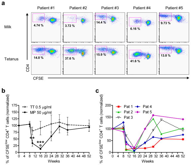
Figure 1. Milk-induced CD4+ T cell proliferation is greatly reduced during the rush desensitization phase.
(a) Frozen PBMC, isolated from 5 milk allergic patients undergoing rush milk desensitization, were thawed and labeled with CFSE, and cultured in the presence of tetanus toxoid (TT) or milk proteins (MP) for 7 days. Cells were then collected, stained with anti-CD4 mAb, and analyzed by flow cytometry. The number in each panel represents the fraction of total CD4+ cells that are within the CFSElow CD4+ gate (antigen-specific proliferating cells) on day 0. Mean CD4+ milk-specific proliferating cells=7.75%; mean CD4+ tetanus specific cells=24.6%. Mean CD4+ proliferating cells without antigen=0.62%.
(b) Each line represents data for all 5 patients combined (solid line, milk protein (MP); dashed line, tetanus toxoid (TT)). Data represent mean % antigen-specific cells (CFSElow CD4+ cells) of total CD4+ cells, normalized to day 0 for each patient ± SEM, over the course of the study. ** p < 0.01, *** p < 0.001 versus baseline and week 8, determined using paired t-test. ° p < 0.05 versus TT response, determined using two-way ANOVA for matched values with Bonferroni’s post hoc test.
(c) Each line represents data for an individual patient, of % milk-specific CFSElow CD4+ cells of total CD4+ cells, normalized to day 0 for each patient, over the course of the study.

