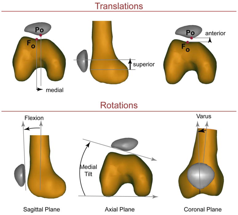
Figure 1. Three-dimensional PF kinematics.
The kinematics reported were based on a 3D anatomical coordinate system established in the patella and femur in the full extension time frame (Seisler and Sheehan, 2007), with medial, superior and anterior shift along with flexion, medial tilt and varus being positive. The kinematics were similarly defined for the tibiofemoral joint with internal rotation being positive. All rotations were calculated based on an xyz-body fixed coordinate system such that the rotation sequence was flexion, tilt, varus rotation.

