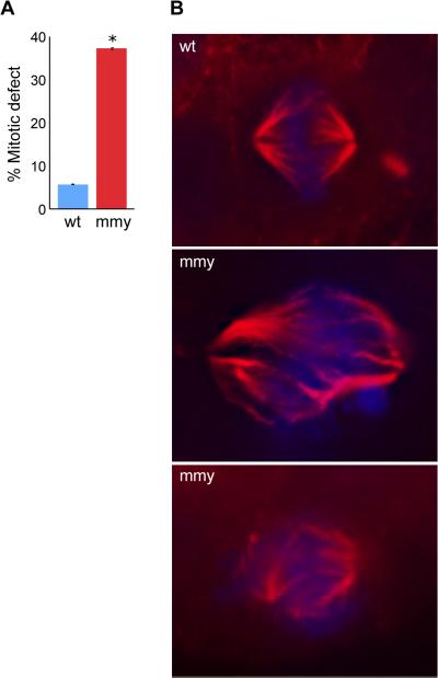
Figure 6. mmy mutant embryos have mitotic defects.
(A) Quantification of immunohistochemistry experiment. Ten wild-type and ten mutant embryos were used to count 180 mitotic events (ME) in wild type and 221 ME in mutant embryos. The embryos were sectioned into head and trunk sections and the total number of ME for each embryo counted. Error bars indicate the standard deviation between each experiment. *: p = 0.000031 (Student's t-Test). (B) Wild-type and mutant embryos were sectioned (10 each), mounted and subjected to immunohistochemistry using an α-tubulin antibody (red) and counter stain for DNA with DAPI (blue).

