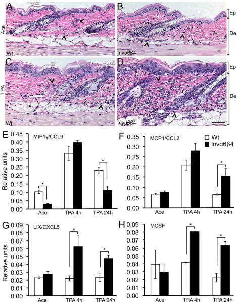
Fig. 1.
Modulated infiltrate and cytokine secretion in TPA-treated Invα6β4 transgenic skin. (A-D) Histological sections from acetone vehicle- (top row) or TPA-treated (bottom row) Wt (left panels) and Invα6β4 (right panels) mouse skin stained with H&E. Arrow heads point to dermal infiltrative cells. Scale bar: 50 μm. Abbreviations: De, dermis; Ep, epidermis. (E-H) Bar graphs depicting the relative units of CCL9, CXCL5, MCP1 or M-CSF compared between acetone vehicle- or TPA-treated Wt and Invα6β4 mouse skin. *- p < 0.05 (Student's t test).

