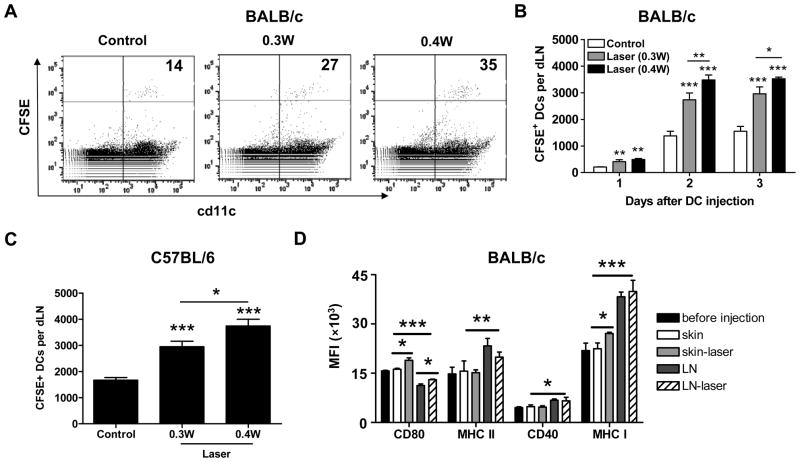
Figure 3. Laser enhances migration and maturation of i.d. injected mDCs.
A–B. Laser enhanced DC migration in BALB/c mice. CFSE-labeled mDCs were i.d. injected to syngeneic BALB/c mice after laser or sham illumination at 0.3W for 2 min or at 0.4W for 1.5 min. Two days later, CFSE+cd11c+ DCs in the dLNs were quantified by flow cytometry and indicated in the upper right corner of the profile (×100) in A or averaged in B. Laser-enhanced earlier (day 1) and later (day 3) DC migration was also similarly quantified as shown in B. C. CFSE-labeled mDCs from B6.SJL mice were i.d. injected to congeneic C57BL/6 (B6) mice and CFSE+cd11c+ cells in the dLNs were calculated on day 2 after DC injection. D. In a second experiment, single cells of the skin and dLNs were immunostained and subjected to flow cytometry analysis of expression of costimulatory molecules (CD80, MHC II, CD40 and MHC I) two days after mDC injection to BAlB/c mice. Mean fluorescence intensity (MFI) of these markers was shown. n=6; *, **, ***, P<0.05, 0.01, 0.001, respectively. Experiments were repeated twice with similar results.

