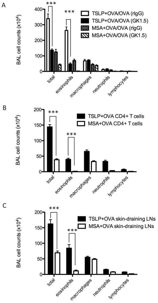
Figure 4.
CD4 T cells are required for TSLP-mediated airway inflammation. (A) Cell counts in BAL fluid from WT BALB/c mice treated i.p. with rIgG or anti-CD4 Ab (GK1.5), to acutely deplete CD4 T cells after skin sensitization. n ≥ 4 mice per group from two independent experiments. (B) DO11.10 CD4+ T cells were transferred to WT BALB/c mice and then sensitized with TSLP+OVA (solid squares) or MSA+OVA (open squares). On D14, CD4+ T cells were isolated and negatively selected prior to transfer of 2 ×10⁷ cells/mouse. Recipient mice were intranasally challenged with OVA starting 24 hours after transfer, and airway symptom were monitored as above. Cell counts in BAL fluid. n = 5 mice per group. The significance between two groups was determined by two-tailed Student’s t test. (C) Skin-draining LN cells from TSLP+OVA treated mice transfer disease to naïve mice. Skin-draining LN cells from mice treated with TSLP+OVA (solid square) and MSA+OVA (open square) were isolated, and cells were cultured with OVA (100 μg/mL) for 72 hours prior to washing and intravenous transfer to naïve mice (2 × 10⁷ cells/mouse). Cell counts in BAL fluid. n = 5 mice per group. The significance between two groups was determined by two-tailed Student’s t test.

