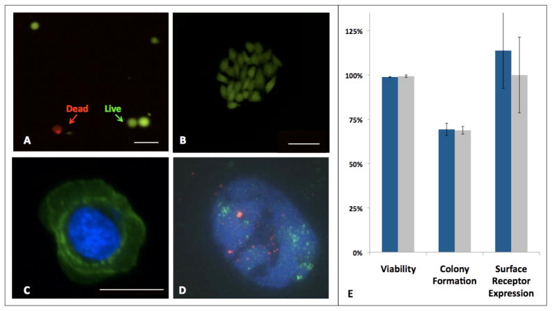
Figure 4.
Released cells were evaluated for (A) viability using a fluorescent LIVE (green)/DEAD (red) assay and (B) colony formation; scale bars are 50 um. Released cells were found to be compatible with downstream (C) immunostaining of cell surface receptors (shown here is HER2 expression in a released cell in green, counterstained with DAPI nuclear staining in blue; 20 um scale bar). (D) FISH analysis, was also feasible as shown here in a released HER2 (green probe) amplified breast cancer cell; the control probe is shown in red. (E) Released cells (blue bars) were found to have comparable viability (98.9% ± 0.3% vs. 99.4% ± 0.6%), rates of colony formation from single cells (69.3% ± 3.4% vs. 68.8% ± 2.2%), and relative surface receptor expression (113.8% ± 21.2% vs. 100% ± 21.3%) when compared to control cells (gray bars) maintained in the appropriate cell culture medium ( p > 0.05).

