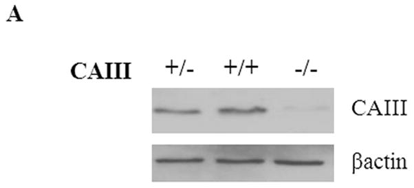
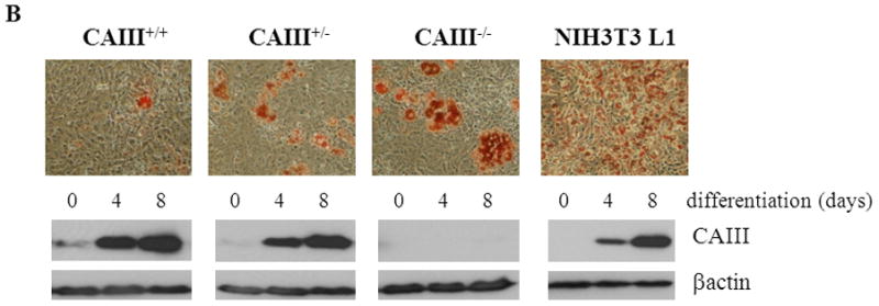
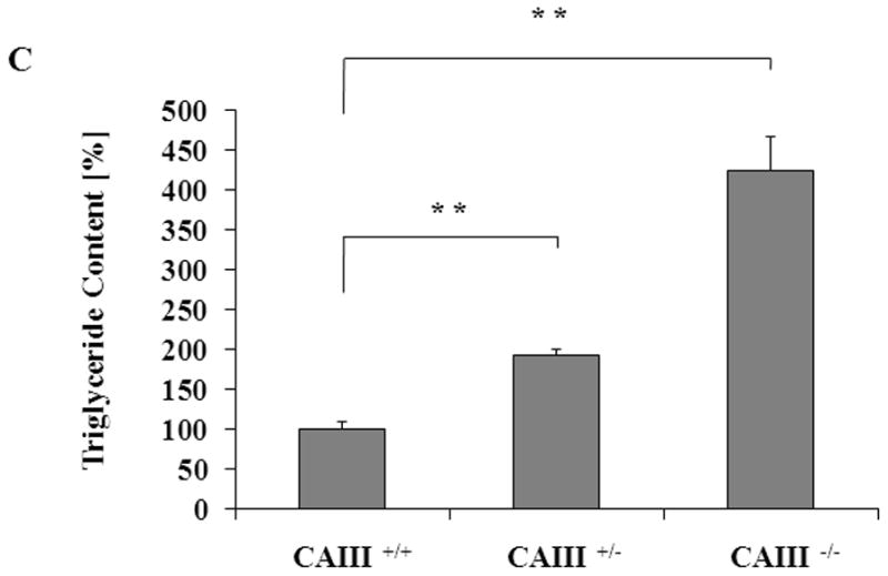
Fig. 1. CAIII knockout enhances fat droplet formation in MEFs.


(A) CAIII protein levels in MEFCAIII+/+, MEFCAIII+/− and MEFCAIII−/− analyzed by Western blotting. β-Actin served as a loading control. (B and C) MEFs were exposed to hormone-cocktail to induce adipogenesis for 8 days. (B) At day 8 of differentiation, the MEFs were fixed, and the fat droplets were stained with Oil Red O (upper panels). Protein samples were taken for western blot analysis at days 0, 4 and 8 (lower panels). Differentiated NIH 3T3-L1 adipocytes are shown as the positive control. (C) The triglycride content in the different MEFs was quantified by densitometric analysis. Data are presented as the mean +/− SD of 4independent experiments, (**) p ≤ 0.01.
