
Figure 7.
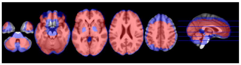
The extent of increased coverage with missing data replacement. Blue voxels were missing data from 18 or fewer subjects and were the focus of data-replacement strategies in the current study. Red voxels show complete voxel-wise data that were submitted to group level analyses under default analysis strategies, while blue would normally be ignored. Whole brain coverage increased by 35.3% following imputation, most notably at the edges of cortex.