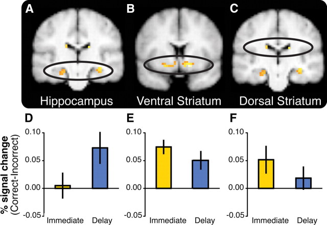
Figure 6.
Activation in the hippocampus and the striatum is differentially sensitive to feedback timing during learning. A–C, BOLD activity in the hippocampus and the striatum correlated with model-derived feedback prediction errors, collapsed across Immediate and Delayed feedback conditions during learning (pSV_FWE < 0.05; maps displayed at p < 0.001 uncorrected within a mask consisting of the striatum and the medial temporal lobe). D–F, ROI analyses revealed that the hippocampus was selectively sensitive to delayed feedback, whereas the striatum was sensitive to immediate feedback. The bar graphs represent feedback sensitivity (BOLD percentage signal change difference between Correct and Incorrect trials) extracted from ROIs (6 mm spheres). D, Left hippocampus [−24 −18 −18]. E, Left ventral striatum [−21 6 −12]. F, Left dorsal striatum [−15 −12 21]. Error bars represent ±1 SEM. Parallel results were obtained in anatomical ROIs.

