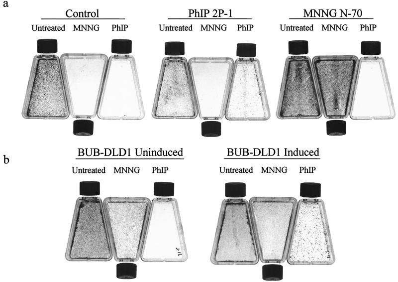
Figure 1.
Resistance to MNNG and PhIP. (a) Approximately 105 cells of control H3 clones (not previously exposed to carcinogens) or clones derived after exposure to carcinogens (see Table 1 for enumeration) were exposed to either 50 μM PhIP or 5 μM MNNG as described in Materials and Methods, and cells were stained with crystal violet 14 days later. Untreated cells served as a plating control (Untreated). (b) BUB-DLD1 cells, inducibly expressing a dominant mutant hBUB1 gene were exposed to PhIP or MNNG and stained as in a. As DLD1 cells are MMR deficient, they are resistant to MNNG, irrespective of induction.

