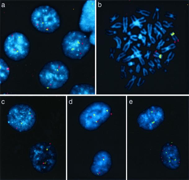
Figure 2.
FISH analysis of chromosomal instability in clones surviving carcinogen exposure. A chromosome 12-specific centromeric probe was labeled with FITC (yellow), and a contig of three bacterial artificial chromosome clones mapping to the distal part of chromosome 12q was labeled with tetramethylrhodamine B isothiocyanate (red). Cells were counterstained with 4′,6-diamidino-2-phenylindole (blue). Nuclei of control cells (a) and MNNG-resistant clones (b and c) exhibited two yellow and two red signals in virtually every nucleus (a and c) and metaphase spread (b). In contrast, cells of PhIP-resistant clones (d and e) often exhibited more or less than two copies of chromosome 12.

