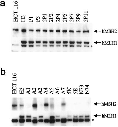
Figure 3.
Expression of hMLH1 and hMSH2 proteins in clones surviving carcinogen exposure. Western blotting was performed with anti-hMLH1 and anti-hMSH2 antibodies. H3 cells expressed full-length forms of both hMLH1 and hMSH2, as did every PhIP-resistant clone (a). In contrast, each MNNG-resistant clone expressed either a full-length hMSH2 or a full-length hMLH1 protein, but not both, demonstrating a highly specific mechanism for loss of MMR gene activity in each clone (b). The asterisk (*) indicates a nonspecific band crossreacting with the anti-hMLH1 antibody.

