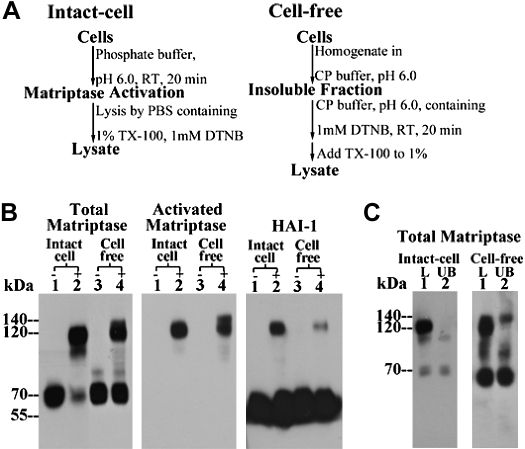
Fig. 3.
Formation of 140-kDa matriptase complex is induced in the cell-free matriptase activation system using 184 A1N4 cells that express high levels of HAI-1. A: flow chart of matriptase activation in the intact-cell and in vitro, cell-free systems. B: less robust matriptase activation in the cell-free activation system compared with that of intact-cell system. Living cells (lanes 1 and 2) or the insoluble fractions (lanes 3 and 4) of cell homogenates of 184 A1N4 mammary epithelial cells were exposed to pH 6.0 buffer alone (lanes 2 and 4, +) or in the presence of 150 mM NaCl as nonactivation control (lanes 1 and 3, −) at room temperature for 20 min. Lysates were prepared and analyzed by immunoblot for total matriptase using mAb M24, for activated matriptase using the mAb M69, and for HAI-1 using the mAb M19, as indicated. C: generation of the 140-kDa matriptase complex in 184 A1N4 cells by in vitro, cell-free matriptase activation method. 184 A1N4 cells were exposed to pH 6.0 buffer to induce matriptase activation in either intact-cell or cell-free settings, as indicated. Lysates were incubated with immobilized HAI-1 mAb M19 to deplete the 120-kDa matriptase-HAI-1 complex. Lysates (L, lane 1) and HAI-1-depleted fractions (UB, lane 2) were analyzed by immunoblot for matriptase species using total matriptase mAb M24. TX-100, Triton X-100; DNTB, 5,5-dithio-bis(2-nitrobenzoic acid); RT, room temperature.

