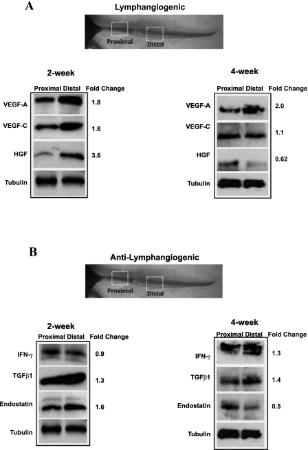
Fig. 3.
Lymphatic fluid stasis regulates expression of lymphangiogenic and anti-lymphangiogenic cytokines. Representative (of triplicate) Western-blot analysis of lymphangiogenic (A) and anti-lymphangiogenic (B) cytokines 2 or 4 wk after surgery comparing expression in regions of the tail located 1 cm proximal to 1 cm distal to the zone of lymphatic obstruction. Gross photograph and boxed regions of the tail are shown for orientation. Fold changes are calculated comparing expression in the distal region to the proximal region after correction for equal loading with tubulin.

