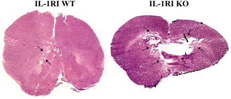
Figure 5. IL-1RI deficiency exacerbates tissue damage during acute brain abscess development.
Representative H&E (haematoxylin and eosin) stained images of brain sections from WT and IL-1RI KO mice at 18 h following S. aureus infection were created by stitching consecutive microscopic fields of view (lesions delineated by arrows). Black areas represent regions devoid of signal during the imaging stitching program. Results are representative of six individual animals per group.

