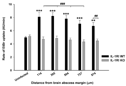
Figure 7. IL-1RI signalling is critical for hemichannel opening during acute brain abscess development.
IL-1RI KO and WT mice received intracerebral injections of S. aureus (103 cfu), whereupon acute brain slices were prepared at day 3 post-infection to monitor hemichannel activity by EtBr uptake assays at the indicated distance from the brain abscess margin (μm). Values represent the means±S.D. from uninfected (six slices, 491 cells analysed) or brain abscess (seven slices, 676 cells analysed) tissues of WT mice, as well as uninfected (six slices, 444 cells analysed) or brain abscess (12 slices, 744 cells analysed) tissues from IL-1RI KO animals. Significant differences between uninfected and infected slices are denoted by asterisks (**P<0.01; ***P<0.001), whereas distinctions between WT and IL-1RI KO slices are indicated by hatched signs (##P<0.01, ###P<0.001).

